
Melanoma

Malignant melanoma is a potentially serious type of skin cancer.
Melanoma Skin Cancer
Malignant melanoma is a potentially serious type of skin cancer. New Zealand has one of the highest incidence rates of melanoma in the world.
Before and After Examples
FAQs
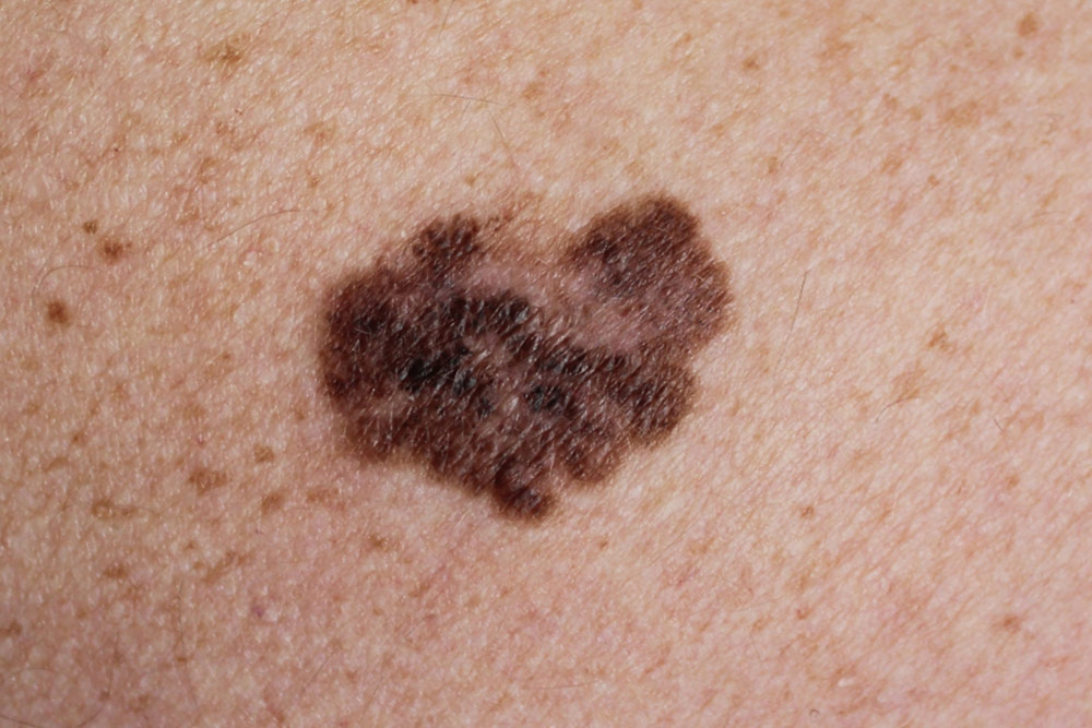
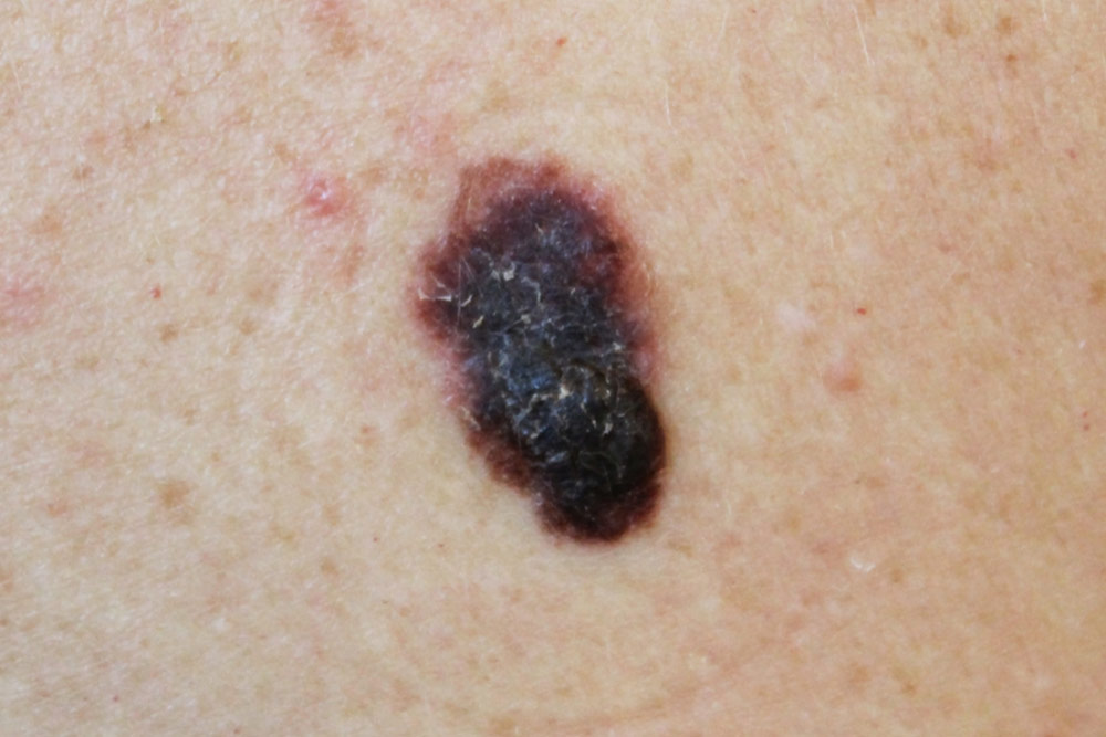
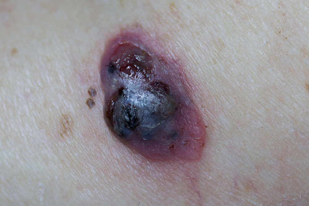
Melanomas arise as a result of the uncontrolled growth of melanocytes (pigment cells) within the skin. They can present as a new mole that looks different to other moles you may have, with features including an irregular border, asymmetrical pattern, colour variation within the mole and/or a larger size. You may also experience symptoms such as itchiness, pain and/or bleeding. These changes can also occur in mole you have had for a long time.
As well as invading the local tissues, melanoma has the potential to spread to the lymph nodes and via the bloodstream. Early diagnosis is key, as it reduces both the complexity of the surgery required to treat it and the risk of melanoma spreading to other parts of your body.
If you are diagnosed with, or suspected of having a melanoma, Dr Collins will discuss the treatment options with you and together formulate your management plan. This may involve a diagnostic biopsy, excision of the scar from a previous biopsy (wide local excision) or wide local excision combined with a sentinel lymph node biopsy.
In certain cases, particularly for thin melanomas, your surgery may comprise of a wide local excision alone. This involves completely excising the melanoma, in addition to an appropriate margin of normal skin, whilst ensuring that scarring is kept to a minimum.
A sentinel node biopsy locates the first lymph node that drains from the area where your melanoma developed. The surgery is performed under general anaesthetic. Before your surgery a radioactive tracer is injected into the scar where the melanoma was removed. A scan is then performed to identify the likely location of the sentinel node and its position is marked on your skin. During the surgery blue dye is also injected into the scar. Both the radioactive tracer and blue dye travel through the lymphatic channels and the first lymph node they reach is removed via a small incision in the overlying skin. The lymph node is examined to determine if there is melanoma present. If it is negative, no further surgery or investigations are necessary. You will, however, need to attend follow-up appointments with Dr Collins for a five-year period.
If there is melanoma present in the lymph node, Dr Collins will arrange for you to have some additional tests such as a CT scan or a PET scan. Depending on a number of factors, you may subsequently be offered further surgery to remove the remaining lymph nodes (lymphadenectomy), or surveillance of the relevant lymph node basin (neck, armpit or groin) with regular clinical examinations and ultrasound scans.
This procedure involves removing all of the lymph nodes in the relevant lymph node basin. It is performed under general anaesthetic. The goal of surgery is to prevent the melanoma recurring in your lymph nodes and to reduce the risk of it spreading to other parts of your body. A drain is inserted at the time of surgery and often remains in place for a few weeks afterwards. The surgery necessitates a 3 to 5 day stay in hospital. Your return to work will be determined by the type of job that you do. If your work is physically demanding, you may require up to 6 weeks of leave.
Attend your follow-up appointments with Dr Collins and have your skin checked regularly. A diagnosis of melanoma means you have a higher than average risk of developing another melanoma, in addition to other types of skin cancer, in the future. Early intervention ensures that any new cancers that develop are diagnosed and treated early and efficiently.